Medical Information
Orthopedic Surgery
- Orthopedic Surgery
- Neurosurgery
- General Surgery
- Pediatrics and Adolescents
- Emergency Medicine
- Kidney Medicine
- Cardiology
- Endocrinology
- Respiratory Medicine
- Gastroenterology
- Family Medicine
- Neurology
- Gynecology
- Dental Clinic
- Anesthesiology and Pain Medicine
- Occupational Environmental Medicine
- Radiology
- Diagnostic Laboratory Medicine
Orthopedic Surgery
Degenerative Arthritis
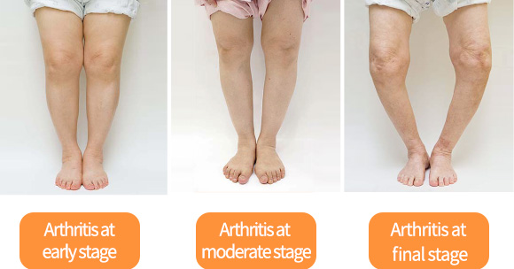
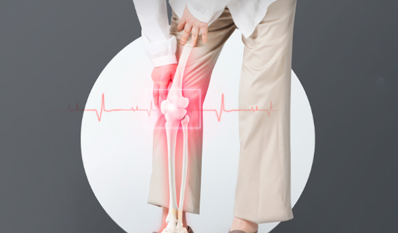
Degenerative arthritis happens when cartilage is worn away or damaged. It causes inflammation and deteriorates the bone. As we age, our joints also experience aging. As joints age, the cartilage wears away and then bones rub on each other, which causes an inflammatory reaction and arthritis. As the most common joint disease, it causes severe pain, difficulty in moving, and even deformation of the joint if unattended for a long time.
| Arthritis Diagnosis | Joint Condition | Symptoms |
|---|---|---|
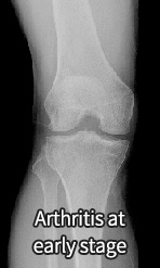 |
Early-stage : minor cartilage damage |
|
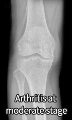 |
Mid-stage : The cartilage wears out and becomes tattered. The ends of the bones grow sharp. |
|
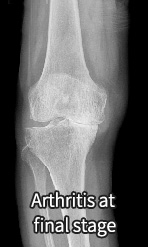 |
Late-stage : The cartilage is extensively damaged and the bones are almost attached to each other. |
|
Treatments of Degenerative Arthritis
Early-Stage Treatment for Arthritis
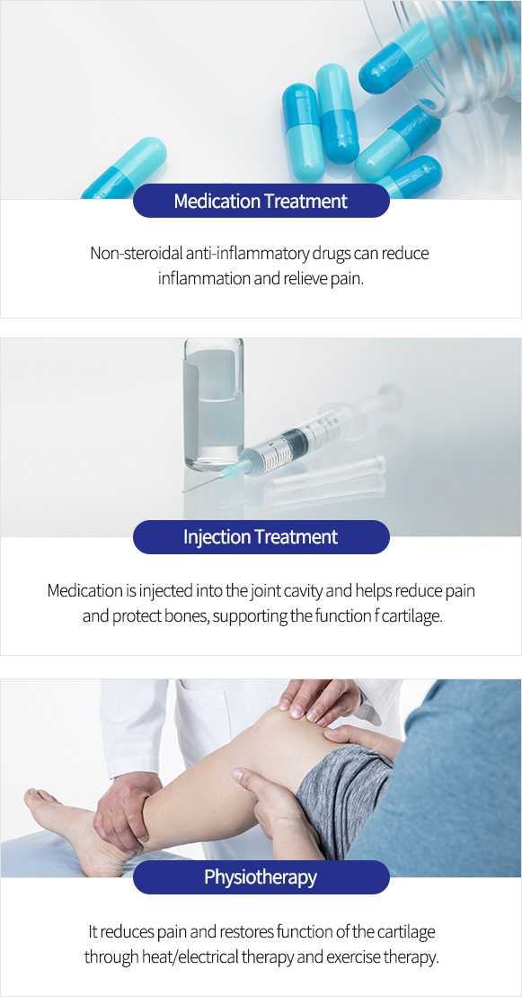
In the early stage when cartilage damage is mild, lifestyle improvement, drug therapy, injection therapy, or exercise therapy can be implemented.
Mid-Stage Treatment for Arthritis
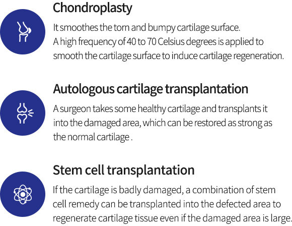
Since the cartilage is not completely damaged or lost, treatments to regenerate the cartilage or to correct the axis of the deformed leg can be applied.
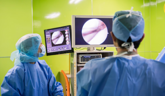
Various approaches are available by inserting an
endoscopic camera into the joint: removing damaged
cartilage, suturing damaged areas or rebuilding the ruptured part.
It is a treatment to correct an O-shaped leg. A surgeon straightens
the leg and gets the weight load to be distributed to the opposite side
without arthritis. When a bent leg is corrected, the weight is shifted
to the outer side, where there is enough cartilage. The cartilage inside
the knee with arthritis can be protected from further wear and tear so that
pain can be reduced and the lifespan of the joint can be extended.
End-Stage Treatment for Arthritis
An artificial joint surgery is the best treatment in the terminal stage when the cartilage is completely worn out.
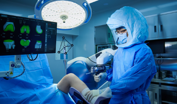
After removing the damaged cartilage, cartilage plate and
ligament, artificial cartilage made of special reinforced
plastic is inserted and it works as a cartilage plate.
Articular cartilage damage
The cartilage acts as a cushion that prevents friction between bones. When this cartilage wears down or out, it causes aching, sore pain. Since the cartilage does not have nerve cells that can make you feel pain, you may not feel any symptoms in a mild case. However, symptoms suddenly get worse before you notice it, so early diagnosis is very critical.
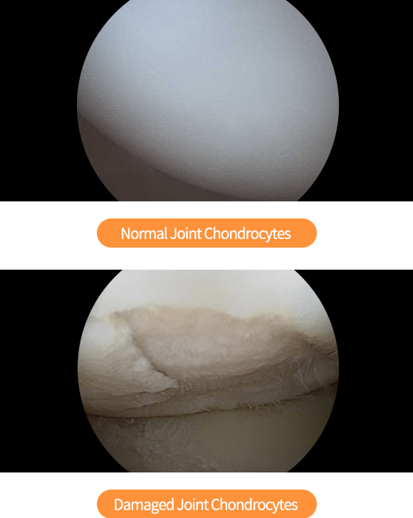
Treatment of Knee Cartilage Damage
-
Micro-drilling
If the size of cartilage damage is less than 1 square cm, an arthroscopy is used to intentionally make a small hole of 3 to 4 mm in the bone under the cartilage so that bleeding and scarring can force the cartilage to regenerate.
-
Autologous cartilage transplantation
If the size of cartilage damage is less than 2-3 square cm, a part of your own healthy cartilage can be taken and transplanted into the damaged area.
-
Autologous cartilage cell culture transplantation
If the size of cartilage damage is wider than 4 square cm, some healthy normal cartilage is collected, cultured, proliferated into cartilage cells and then transplanted into the defected area.
Meniscus Injury
The meniscus is a crescent-shaped cartilage plate located between thigh bone and calf bone. It reduces the impact on the joint and helps the joint to move freely. A tear or rupture of the meniscus is called a meniscus injury. Depending on the type of rupture, various types of damage can be classified.
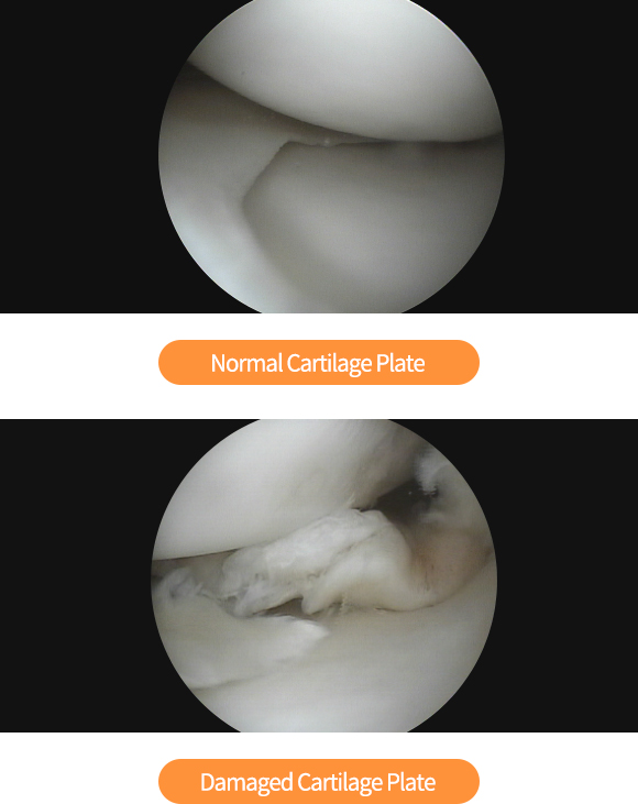
Meniscus cartilage damage
There are treatments including a meniscal resection to trim the torn area using an arthroscopy, a meniscus suture, and a meniscal cartilage graft to transplant a new one.
-
meniscus suture
It is a treatment in which a surgeon sutures the ruptured cartilage plate. The operation result is good when a patient is young or the injury happened from traumatic shock.
-
Meniscal resection
This treatment can be applied when the cartilage plate does not adhere even after sutured or when it is difficult to suture. If the rupture is left unattended, the damage can be extended so the ruptured part should be excised.
-
Meniscal cartilage graft
In this treatment, a specially-treated living cartilage plate is implanted. As the cartilage plate is a bloodless tissue, engraftment is available without rejection reaction.
Knee ligament injury
The knee has a collateral ligament that firmly connects the left and right side, and a cruciate ligament that allows the knee to move along a planned trajectory without swaying back and forth. They are very flexible and can manage damage caused by impact or rotational force. But their elasticity has a limit like a rubber band and can be torn when they go beyond the limit.
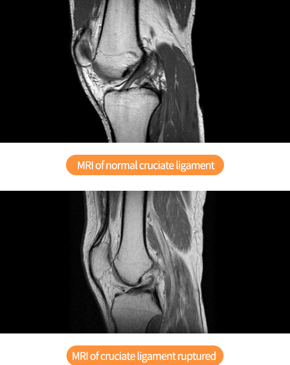
Treatment of Knee Cruciate Ligament Injuries
-
Conservative treatment
When the cruciate ligament damage is minor without functional problems, exercise therapy can be performed after being stabilized for 4 to 6 weeks, using an orthosis or crutches without weight bearing.
-
Ligament reconstruction
If the rupture is not serious, sutures to connect the torn ligaments are performed. But when the cruciate ligament is completely ruptured, it can be reconstructed in two ways: rebuilding the ligament with another tendon in the knee or transplanting tissues of other people.
* In rare cases, bleeding, infection, and blood clots may occur during surgery.


